Canine Schistosomiasis
August 4, 2026 · Internal Medicine

History: The patient was presented to the internal medicine service at GCVS NASA Parkway for a history of recurrent hematochezia (bloody stools), vomiting, and inappetence. The patient’s symptoms began about a year prior to his presentation, with little to no real improvement noted after several diet trials and courses of various antibiotics and probiotics. Comprehensive bloodwork and fecal testing was eventually performed, documenting an increase in his total protein (9.4) and globulins (6.4) as well as a low cholesterol (92). Because of his poor response to treatment, the patient was eventually placed on a course of steroids (prednisone), which resulted in near resolution of his clinical signs. Over the next 6 months, multiple attempts to wean him from prednisone led to a recurrence of his clinical signs for which further consultation was sought. The patient was a strictly indoor dog with no recent travel history. He was current on all vaccinations and was receiving treatment with psyllium husk, prednisone, and FortiFlora.
Examination: On examination by our service, the patient appeared bright, alert, and otherwise in good spirits. He was adequately hydrated with pink mucous membranes and normal vital parameters. He was mildly overweight with a pot-bellied appearance and bilaterally symmetrical alopecia (hair loss). Auscultation of his chest revealed otherwise clear lung sounds in all fields with no evidence of heart murmur or arrhythmia. His abdomen was soft and non-painful with mild liver enlargement and subjective bowel thickening noted. Digital rectal exam was unremarkable, and evidence of peripheral lymph node enlargement was not appreciated.
Diagnostic Testing performed with IM: Given his young age and concern for either a primary endocrine and/or gastrointestinal disorder an initial set of diagnostics to include comprehensive a comprehensive CBC and chemistry panel, GI profile, ACTH stimulation test, histoplasma and heterobilharzia testing, thoracic radiographs, and abdominal ultrasound was performed. Results are provided below:
CBC/chemistry panel: increased total protein (9.2 g/dL) and globulins (5.8g/dL) with a low potassium (3.2mmol/L)
ACTH stimulation test: resting 1.4mcg/dL, post 4.5mcg/dL
GI profile: normal TLI/PLI and cobalamin and folic acid levels
Histoplasma antigen: negative
Abdominal ultrasound: Diffusely thickened small intestinal submucosal layer with duodenal hyperechoic mucosal striations and colonic mucosal thickening. Mild gastroparesis. Bilaterally small adrenal glands.
Thoracic radiographs: normal thorax
Heterobilharzia PCR: positive
Treatment and Follow-up: Pending the results of his GI profile, histoplasma antigen, and heterobilharzia PCR tests, it was recommended that the patient be placed on a lower dose of prednisone in the hopes of weaning this medication while also being maintained on his psyllium husk and fortiflora. Upon confirmation of positive heterobilharzia test, he was started on a 10-day course of fenbendazole (40mg/kg) given once daily and a 2-day course of praziquantel (15mg/kg) given every 8 hours. This course was repeated a month later, at which time he was found to be doing very well at home with half a pound of weight gain and no vomiting, diarrhea, or inappetence noted. His prednisone and psyllium husk was also discontinued at that point. A month after completing his second course of fenbendazole and praziquantel, the patient was again reported to be doing very well, with another quarter of a pound of weight gain noted. Repeat heterobilharzia was performed at that time and was subsequently found to be negative, confirming clearance of his infection.
Discussion: American schistosomiasis, also known as a heterobilharzia infection, is caused by Heterobilharzia americana, a trematode (flatworm) parasite that primarily affects the liver and intestinal blood vessels of dogs. Infection occurs when dogs are exposed to freshwater environments containing infected freshwater snails, which serve as an intermediate host. After entering the dog, the parasite migrates through the bloodstream over a period of 2-3 months, gradually maturing into an adult. They eventually settle in the liver, intestine, and other organs, where they begin shedding eggs, resulting in significant inflammation and organ damage. Clinical signs can vary from mild gastrointestinal upset to more severe disease, including weight loss, vomiting, diarrhea, lethargy, abdominal discomfort, and liver dysfunction. Some dogs may develop elevated liver enzymes and protein abnormalities, while others can develop chronic complications such as cancer and immune-mediated conditions. Diagnosis can be challenging given the non-specific nature of the disease but usually can readily be made by observing very unique pathologic changes to the small intestine on abdominal ultrasound and detecting the parasites eggs via specialized PCR testing on feces.
Treatment typically involves the use of one or more anti-parasitic medications combined with supportive care, and early recognition is important to reduce organ damage. Treatment is generally very well tolerated, and most animals begin showing clinical improvement within 1-2 weeks of initiating therapy. With proper treatment, the overall prognosis is very good, with few animals experiencing long-term sequelae from their infection. Because the parasite requires freshwater snails for its life cycle, prevention focuses on limiting exposure to contaminated water sources, especially in endemic regions such as parts of the southeastern United States.
This patient’s case is a perfect example of how heterobilharzia infections can readily be confused for other problems and underscores the importance that comprehensive testing can have on proper treatment.
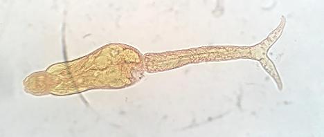
Heterobilharzia americana
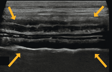
Ultrasonographic image showing severe thickening of the small intestinal submucosa with numerous submucosal and adjacent mucosal punctate hyperechoic foci (arrows).
Partner with our internal medicine team for complex GI workups or explore more case studies from GCVS.
Author: Paul Manino, DVM, DACVIM