Duke: Necrotizing Fasciitis
Signalment:
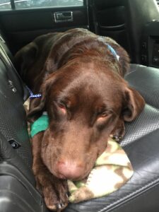
1 year old male neutered Labrador Retriever
History:
Duke was neutered by his primary care veterinarian and had a testicle that was in the inguinal canal that had not fully descended. After surgery, Duke did not fully recover and there was progressive swelling at the surgical site. Duke continued not to feel well, and the swelling progressed up along his body wall. Hemorrhage was noted at the surgery site and bloodwork revealed prolonged blood clotting, requiring blood and plasma transfusions. Due to his need for continued aggressive treatment and progressively critical state, Duke was referred to Gulf Coast Veterinary Specialists.
Physical examination:
Quiet, dull, and responsive. Previous neuter site was open and there was purulent discharge. There was palpable pocketing along the body wall extending from the left groin to the level of the right axilla that was warm and painful to the touch. There was bruising of the skin extending from the inguinal region to the cranial extent of the swelling that was rapidly progressive throughout evaluation period. Duke had poor pulse quality and appeared approximately 7% dehydrated.
Diagnostics:
Complete Blood Count:
- Leukopenia (low white blood cell count)
Biochemical Profile:
Fluid analysis from body wall swelling:
- Beta-hemolytic streptococcus grew on aerobic culture
Abdominal Ultrasound:
- Extensive ventral body wall cellulitis and subcutaneous emphysema with regional lymphoid hyperplasia. Right ventral abdominal wall panniculitis infected with gas producing bacteria.
Surgical Plan:
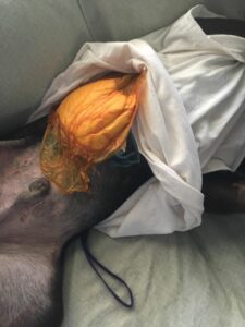
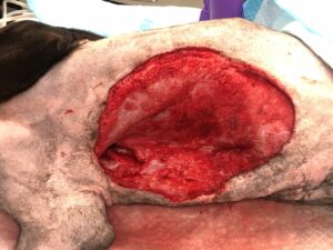
- Debridement of inguinal wound dehiscence
- Drainage of thoracic and abdominal wall abscess and debridement of large amounts of necrotic tissue extending from the right axillary (armpit) region to the left inguinal wound
- Necrosis extending from skin to deep fascia was noted through the majority of pectoral, thoracic wall, and external abdominal muscle groups
- Negative pressure wound therapy device placed over right body wall at -125 mm Hg for 4 days followed by open wound management with delayed wound closure 1 week after initial wound treatment
- Hyperbaric oxygen therapy at 2 atmospheres during open wound management period
- Repeated 2 weeks after wound closure due to development of antimicrobial-resistant Enterobacter infection and partial wound dehiscence
Supportive Care:
- Continued negative pressure wound therapy device
- Nutritional support
- Culture and sensitivity-guided antibiotic therapy
- Multi-modal analgesic therapy
Pathophysiology:
Necrotizing fasciitis
- Necrotizing fasciitis (NF) is a rapidly progressive necrotizing soft-tissue infection that spreads along the subcutaneous tissue accompanied by necrosis of fascia and underlying muscle
- Necrotizing fasciitis (NF) is a rapidly progressive necrotizing soft-tissue infection that spreads along the subcutaneous tissue accompanied by necrosis of fascia and underlying muscle
- Delay in surgery of more than 24 hours was correlated with a 4-fold increased relative risk of mortality
- Dissecting gas or gas opacities in the subcutaneous tissue can been seen on radiographs; this is diagnostic for necrotizing fasciitis in the absence of trauma as was seen on Duke’s ultrasound
- Initial debridement is essential and is performed to completely excise affected tissue. More aggressive debridement resulted in a 43% mortality rate versus 71% with conservative debridement to salvage tissue or a limb
- While a single aggressive surgical resection can be curative, repeat procedures are typically required. The average number of procedures performed in affected humans is approximately three
Hyperbaric oxygen therapy (HBO2):
- Hyperbaric oxygen therapy involves placing the patient in an environment of increased ambient pressure while breathing 100% oxygen for a short period of time, resulting in enhanced oxygenation of the arterial blood and tissues
- For anaerobic infections, the tissue oxygen tensions generated will be bacteriostatic and halt production of particular bacteria
- Infected tissue is known to be hypoxic through a combination of poor perfusion and edema, while HBO2 therapy can improve neutrophil function by raising the oxygen tension within the infected tissue
- Hypoxia may also reduce the effectiveness of several antibiotics (vancomycin hydrochloride and ciprofloxacin hydrochloride) while hyperoxia may potentiate the action of others (aminoglycosides cross the cell membrane of the microorganism by an oxygen-dependent pump)
- Once the infection has been controlled, fibroblast stimulation by HBO2 therapy may assist with wound closure
- The incorporation of HBO2 therapy in the management of NSTI was associated with almost 9 times increased survival in a human retrospective study
- overall mortality in the HBO2 group was 23% of 148 patients compared with 48% of 88 patients in the non-HBO2 group
Negative Pressure Wound Therapy:
- Therapy with A.C. has been shown to provide wound-healing properties in regard to:
- Antimicrobial properties via mechanical and oxygen tension properties
- Differentiation of mesenchymal cells into myofibroblasts
- Promotes cellular proliferation
- Promotes neovascularization
- Speeds up the plasmatic imbibition and inosculation phases of free graft healing
- Indications for use:
- Acute and traumatic wounds
- Chronic open wounds without active necrosis
- Skin flaps
- Meshed free grafts
- Pressure/decubital ulcers
- Contraindications
- Necrotic tissue w/out debridement
- Untreated osteomyelitis
- Exposed nerves and/or major vessels not protected with non-adherent bandage material
- Tumor malignancy
Outcome:
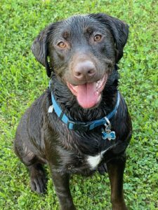
Duke was hospitalized for a week and a half after initial wound debridement and V.A.C. placement. After the negative pressure wound device was removed and the wound managed open until it could be adequately sutured together, Duke was discharged. Two weeks after discharge a small portion of the incision dehisced, and Duke was re-evaluated and wound revision. He went on to heal without complication and is currently running around with his sister and refusing to get out of the pool!
Further Reading:
- Maguire P, Azagrar J M, Carb A & Lesser A (2015) The successful use of negative-pressure wound therapy in two cases of canine necrotizing fasciitis. JAAHA 51 (1), 43-48. https://doi.org/10.5326/JAAHA-MS-6033
- Greijdanus-van der Putten, Vos J T, Devekot J R V et al (2014) [Postvaccinal fatal Streptococcus zooepidemicus necrotizing fasciitis in a young dog: a case report]. Tijdschr Diergeneeskd 139 (9), 24-27 https://pubmed.ncbi.nlm.nih.gov/25272902/
- Bowlt K L, Pivetta M, Kussy F, Rossanese M, Stabile F, Dennis R & Holloway A (2013) Imaging, diagnosis and minimally-invasive management of necrotizing fasciitis in a dog. Vet Comp Ortho Traumatol 26 (4), 323-327 https://doi.org/10.3415/vcot-12-08-0100
- Miller W H, Griffin C E & Campbell K L (2013) Streptococcal and staphylococcal toxic shock and necrotizing fasciitis. In: Muller & Kirks Small Animal Dermatology 7th Edition. pp 214-215.
- Levett, Denny Z, et al. “Adjunctive Hyperbaric Oxygen for Necrotizing Fasciitis.” Cochrane Database of Systematic Reviews, 2011.
- Mastrocco, Alicia, and Jennifer Prittie. “Early and Aggressive Surgical Debridement and Negative Pressure Wound Therapy to Treat Necrotizing Fasciitis in Three Dogs.” Veterinary Surgery, 2021. https://doi.org/10.1111/vsu.13576
- Wilkinson, David. “Hyperbaric Oxygen Treatment and Survival From Necrotizing Soft Tissue Infection.” Archives of Surgery, vol. 139, no. 12, 2004, p. 1339. https://doi.org/10.1001/archsurg.139.12.1339